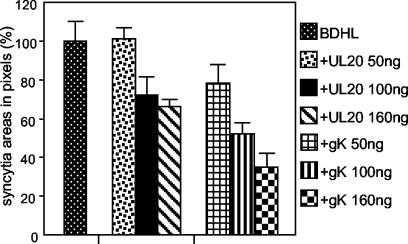
FIG. 2.
Quantification of cell-cell fusion in BHK cells cotransfected with plasmids encoding only gB, gD, gH, and gL (BDHL) (40 ng of each) or gB, gD, gH, and gL plus pUL20-pcDNA or pgK at the indicated amounts and the pcDNA 3.1(−) Myc-His/Lac vector (80 ng). The amounts of DNA in the transfection mixtures were made equal by addition of pMTS1, as appropriate. Syncytia were stained with X-Gal at 48 h. A digital micrograph of the entire coverslip was taken. The blue areas corresponding to syncytia were quantified with the Histogram software of Photoshop. Samples were run three times. The bars indicate standard errors (SE).

