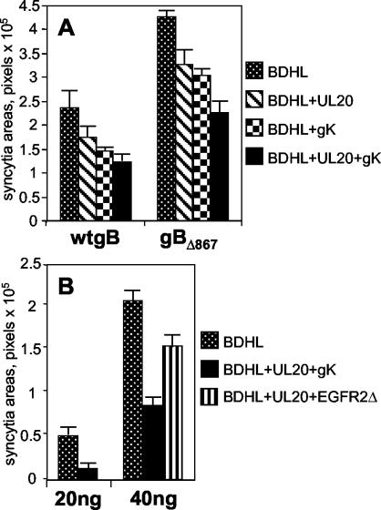
FIG. 4.
(A) Quantification of cell-cell fusion in COS cells cotransfected for 48 h with expression plasmids encoding wt gB, gD, gH, and gL (40 ng of each) (wtgB) or gBΔ867, gD, gH, and gL (20 ng of each) (gBΔ867) plus pUL20-pcDNA and/or pgK (160 ng each). (B) Quantification of cell-cell fusion in COS cells cotransfected for 48 h with expression plasmids encoding wt gB, gD, gH, and gL (20 or 40 ng of each) plus fourfold-higher amounts of pUL20-pcDNA and pgK or EGFR2Δ. BDHL, gB, gD, gH, and gL. The transfection mixtures contained the pcDNA 3.1(−) Myc-His/Lac vector (40 ng), and amounts of DNA in the transfection mixtures were made equal by addition of pMTS1, as appropriate. Syncytium staining and quantification were performed as described in the legend to Fig. 2. The bars indicate SE.

