FIG. 6.
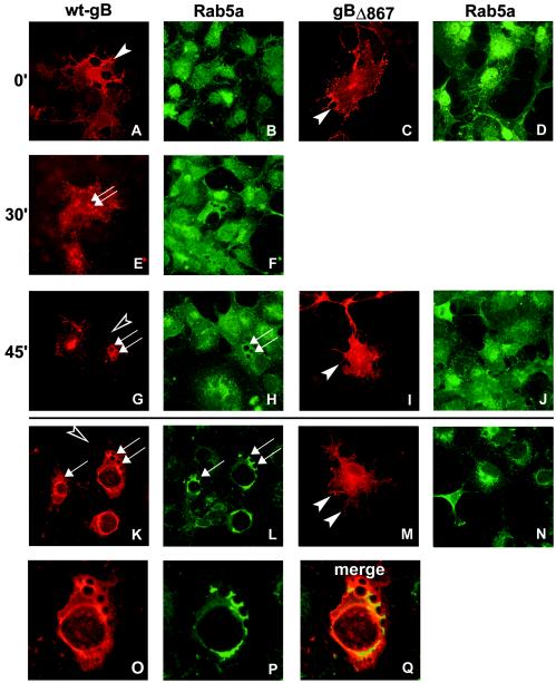
Micrographs of COS cells transfected with pgB-MTS (encoding wt gB) (A, B, E, F, G, H, K, L, O, P, and Q) or pgBΔ867-MTS (encoding gBΔ867) (C, D, I, J, M, and N) and reacted with MAb to gB (A, C, E, G, I, K, M, and O) or MAb to Rab5a (B, D, F, H, J, L, N, and P). (A to J) Endocytosis assays. Cells were reacted with MAb to gB at 4°C and then shifted to 37°C for the indicated times (in minutes), fixed, permeabilized, and reacted with secondary antibodies. Arrows indicate vesicles lined with gB and Rab5a. Filled arrowheads point to gB-labeled cell surfaces. Open arrowheads point to a lack of gB on cell surfaces. (O and P) Higher magnification of panels K and L, respectively. (Q) Merged image of panels O and P shows colocalization of Rab5a and gB.

