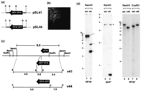
FIG. 1.
(a) Schematic diagram of the transfer plasmids pSL41 and pSL44 which contain the genes for VP16-GFP and GFP-VP16, respectively, in each case flanked by 200 bp of the 5′ and 3′ regions of the native VP16 gene, constructed as described in Materials and Methods. These plasmids were cotransfected with purified HSV-1 [17] DNA, and autofluorescent plaques (b) were isolated and plaque purified. (c) A schematic representation of the VP16 region and the native BamHI and EcoRV sites is illustrated, together with the predicted structure after recombination into the locus. (d) Diagnostic Southern blotting of DNA from stocks of plaque-purified virus was performed to confirm recombination. DNA samples from HSV wt, v41, or v44 viruses are indicated at the top of each lane together with the diagnostic enzyme, and the radiolabeled probes, either VP16 or GFP, are indicated at the bottom. The profile of fragments precisely reflects that expected in panel c. The band marked with an asterisk is likely due to a weak hybridization because of the presence of a short region from HSV thymidine kinase gene in the commercial vector (pEGFPN) used to make the GFP probe.

