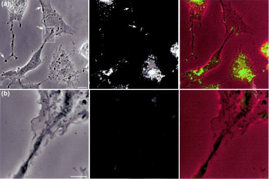
FIG. 11.
HSV infection induces long extensions along which VP16-GFP clusters accumulate. The demonstration of long cellular projections induced by HSV infection is best seen when cells are plated at a relatively low density. Vero cells plated at low density were infected with HSV-1 v41 as before and analyzed by phase microscopy and for VP16-GFP localization. In the combined images (right-hand panel) the phase image is placed in the red channel of the color image, while the VP16-GFP signal is in the green channel. (a) In the top panel long extensions can be seen which can have a bifurcated or bulbous end (arrow), while the right-hand cell shows a similar extension impinging upon a cell not yet expressing VP16. The arrows point to VP16-GFP material which overlies this uninfected cell but originates from the lower infected cell in the panel. Bar, 10 μm. (b) Enlargement of the boxed section of panel a, as discussed in the text. Bar, 5 μm.

