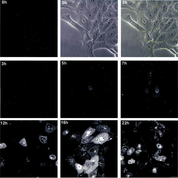
FIG. 4.
Localization of VP16-GFP in virus infected cells. Vero cells, grown in chambered coverslips were infected with HSV-1 v41 at an MOI of 10 for 1 h, washed in buffer, and reincubated at 37°C in medium containing 2% calf serum. At various times thereafter the cultures were removed from the incubator and images captured in live cells as described in Materials and Methods. Representative images are show wherein the extended depth of field was captured by Z-sectioning through the depth of the cell and then stacking the sections using the appropriate Zeiss LSM software module. The time point at 0 h (i.e., 1 h after addition of virus), was imaged both for GFP and for phase contrast, both fields being combined in the top right hand panel. Bar; 25 μm.

