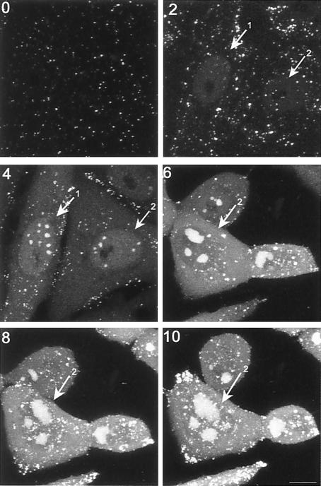
FIG. 5.
Progression of localization of GFP-VP16 in individual infected cells. Vero cells grown on gridded coverslips so that individual specific cells could be tracked, were infected with HSV v44 at an MOI of 10. The cells were infected and replaced in the incubator, and live cell images were recorded using the same settings and laser attenuation at various times thereafter (time indicated in hours postinfection on each panel). This time course highlights key aspects of the nuclear phases of localization where VP16 first appears in a general diffuse pattern, concentrating in subnuclear foci which fuse into large compartments, and additionally is recruited to numerous smaller foci which begin to form around 8 to 10 h after infection. Note that the imaged field for the 6, 8, and 10 h time points had shifted slightly to the right. Bar, 10 μm.

