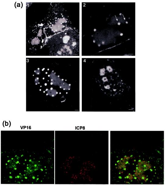
FIG. 6.
GFP-VP16 localization in replication compartments. (a) Aspects of GFP-VP16 localization in the nucleus were studied in more detail using Z-sectioning and higher magnification imaging. Vero cells were infected and live cells imaged as described above. In panel 1 distinct small foci directly impinging on the replication compartments can be readily observed (arrows). They are spherical and relatively homogeneous in size distribution. Occasionally (panel 2), it appeared that these novel foci had themselves substructures resembling coalescing microfoci, but this was generally difficult to discern. The majority of the smaller GFP-VP16 foci appeared to be associated with the perimeter of the larger replication foci (panel 3). By sectioning cells through the larger foci, areas devoid of GFP-VP16 could be identified, as shown in panel 4. Bar; panel 1, 5 μm; panels 2 to 4, 2 μm. (b) VP16 colocalizes with ICP8 in replication compartments, but ICP8 is not recruited into the adjacent foci. To confirm that the larger coalesced compartments containing GFP-VP16 represented replication compartments, infected cells were fixed and stained with antibody to ICP8 or to GFP (as indicated). With this antibody ICP8 is observed in a microspeckled pattern in the compartments, and there was clear colocalization with VP16. However, ICP8 was absent from the VP16-containing foci which formed on the perimeter of the replication foci. Bar, 2 μm.

