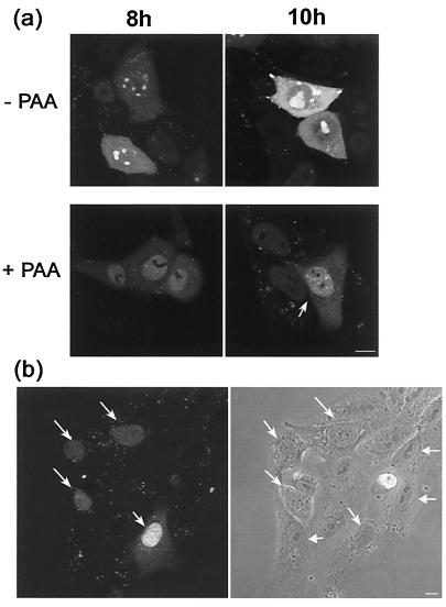
FIG. 7.
Effect of inhibition on DNA replication on localization of GFP-VP16. (a) Vero cells plated in chambered coverslips were infected with HSV-1 v44 at an MOI of 10, the inoculum replaced 1 h later and the cells further incubated in the presence or absence of PAA at a concentration of 400 μg/ml as indicated. At various time points (indicated in hours) the localization of GFP-VP16 was examined by confocal microscopy in live cells as discussed in the text. (b) Disruption of normal nucleolar organization early in infection. Vero cells infected with HSV-1 v44 at an MOI of 10 analyzed very early after infection (2 h) show de novo-synthesized VP16 in a diffuse nuclear pattern (arrows). When examined by phase microscopy these same cells show a distinctly altered gross nucleolar morphology comprising an untangled fibrillar type pattern (diagonal arrows) readily distinct from the more compact, phase dense lobular morphology in those cells not yet expressing VP16 (horizontal arrows). Bar, 10 μm.

