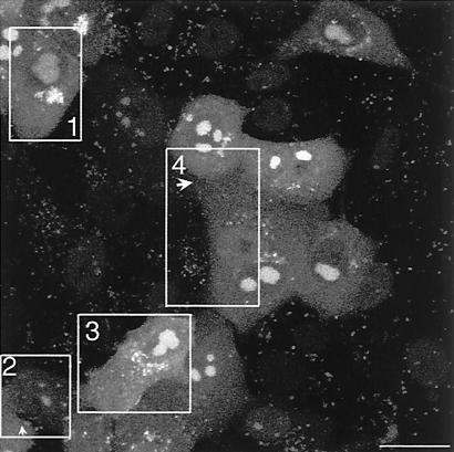
FIG. 9.
Analysis of VP16-GFP movement and accumulation within infected cells by time lapse. This figure represents the first frame of a QuickTime movie. Vero cells were plated on 40-mm coverslips which were assembled into a Bachhoffer POC-chamber (Carl Zeiss Ltd.) which could be maintained at constant temperature and with a controlled CO2 environment on the confocal microscope. Cells were imaged using the XYZT module of the LSM410 imaging software, collecting 10 individual sections separated by 1.5 μm at each time point, stacking the sections to give one extended depth of focus and compiling the individual images into a QuickTime movie series. Several movies were compiled. This series summarizing some of the key features discussed in the text was taken at 72 h after an original infection at an MOI of 0.001. Each of 150 frames was taken at 5 min covering a total time span of 12 h. Bar, 50 μm.

