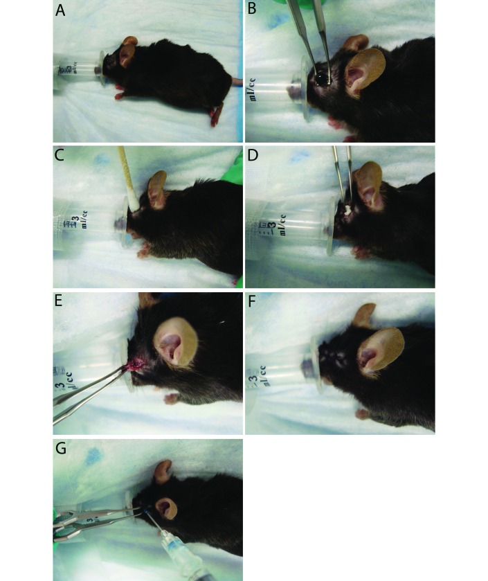
Figure 1.
Enucleation procedure. (A) An anesthetic nose cone was created by removing the top of a 3-mL syringe. This replaced the standard nose cone, allowing for adequate access to the eye while maintaining a surgical plane of anesthesia. Each mouse received a preoperative dose of buprenorphine at induction. (B) Once a surgical plane of anesthesia was obtained, the periorbital region was shaved. The globe was proptosed and isolated with Iris forceps. The globe was removed (without ligation of vessels), and the orbit was flushed with sterile saline. (C) A sterile swab was used to provide hemostasis until active bleeding had ceased. (D) The orbit was packed tightly with absorbable gelatin sponge. (E) The eyelid margins were trimmed to provide fresh tissue for closure. (F) The site was closed using a simple interrupted pattern of 4-0 polydioxanone suture. (G) A drop of skin glue was added to the site to prevent dehiscence due to self-trauma.

