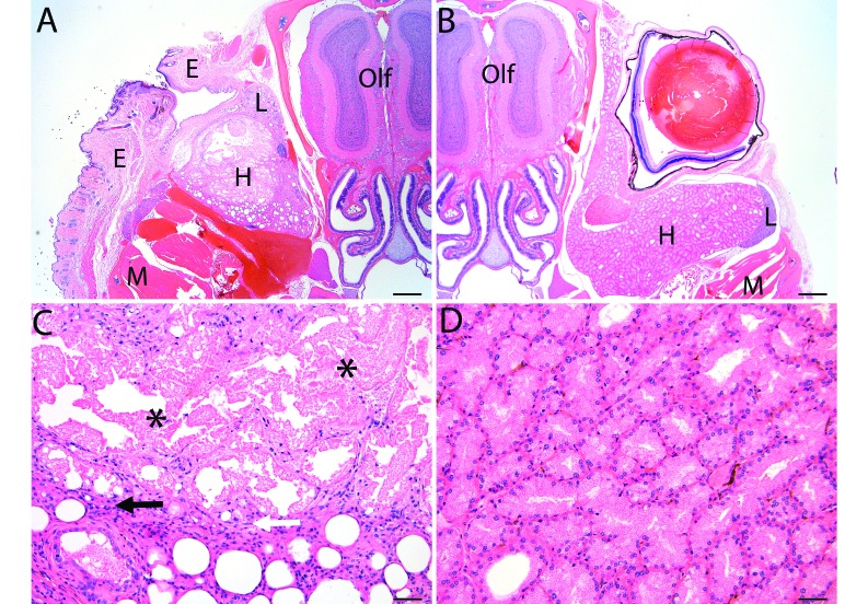
Figure 2.
Histologic evaluation of (A) the enucleation site at 10 wk postoperatively compared with (B) contralateral control. Necrosis and mild granulation tissue occurred within the eyelid at the site of enucleation, but no evidence of infection, such as suppurative inflammation or bacteria, was present. Original , 20×; scale bar, 500 μm. Higher magnification of the enucleation site revealed necrosis (asterisks) with acinar dilation or atrophy in the Harderian gland, which was occasionally accompanied by mild granulomatous inflammation (arrows) that did not extend into the surrounding soft tissue. E, eyelid; H, Harderian gland; L, lacrimal gland; M, muscle; Olf, olfactory lobe of brain. Scale bar, 50 μm.

