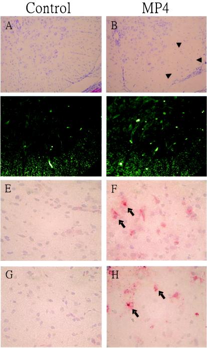
FIG. 6.
Neuronal loss, apoptosis, and VP-1 expression in EV71-infected neural tissues. Seven-day-old mice were orally inoculated with cell lysate (mock control) or the mouse-adapted EV71 strain (MP4) as described in the legend to Fig. 5. Representative sections are shown, demonstrating neuronal loss (indicated by arrowheads) and apoptosis as detected in the ventral horns of the thoracic spinal cord by Nissl staining (A and B) and TUNEL (C and D), respectively, following inoculation with EV71. The immunohistochemical expression and localization of VP-1 (indicated by arrows) are shown for tissues from the brains (E and F) and spinal cords (G and H) of control and MP4-infected mice. Magnifications, ×100 (A to D) and ×400 (E to H).

