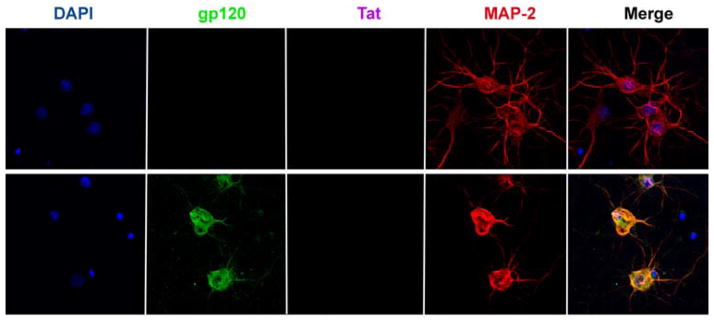
Figure 2. HIV sheds gp120.

Rat cortical neurons were exposed to (upper panel) heat inactivated HIV or (lower panel) T-lymphotropic HIV (AIDS Research and Reference Reagent program, NIAID, NIH, Bethesda, MD) for 24 h. Neurons were then fixed and stained for gp120, Tat and MAP-2. Please note that Tat was undetectable and that neurons exposed to gp120 exhibit shorter processes.
