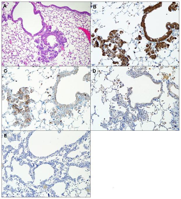
Figure 7.
A H/E-stained invasive SCC of airway (A), and immunohistochemical staining of the same airway for CK5/6 (B), p63 (C), TTF-1 (D), and Napsin-A (E). CK5/6 and p63 staining is positive, while TTF-1 and Napsin-A staining is negative, indicating squamous cell origin of dysplastic tissue. magnification: A. – 100X, B.–E. – 200X.

