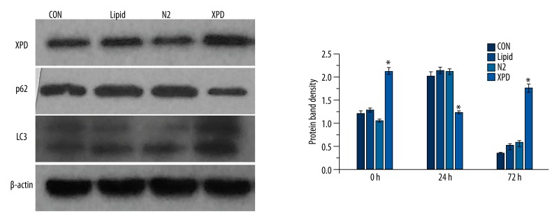
Figure 4.
Western blotting. XPD expression in the XPD group was significantly increased (* p<0.05). Notably, in the XPD group, LC3 expression was significant increased (* p<0.05), while p62 expression was significantly reduced (* p<0.05), as compared to the three control groups. Among the three control groups, differences in protein expression were not statistically significant (p>0.05).

