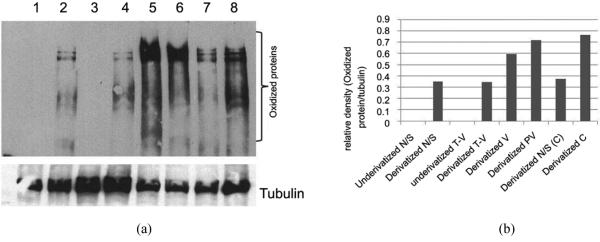
Figure 3.

(a) Western blot analysis of oxidized protein in cytosolic fraction of colorectal adenopolyp tissue. Lane 1, underivatized lysate of normal tissue; Lane 2, DNPH-derivatized lysate of normal tissue; Lane 3, underivatized lysate of tubular villous tissue; Lane 4, DNPH-derivatized lysate of tubular villous tissue; Lane 5, DNPH-derivatized lysate of villous tissue; Lane 6, DNPH-derivatized lysate of polypvillous tissue; Lane 7, DNPH-derivatized lysate of normal tissue surrounding carcinoma tumor; Lane 8, DNPH-derivatized lysate of carcinoma tissue; (b) The relative optical density on the level of reactive carbonyl proteins using myImageAnalysis v 1.1, Thermo Scientist.
