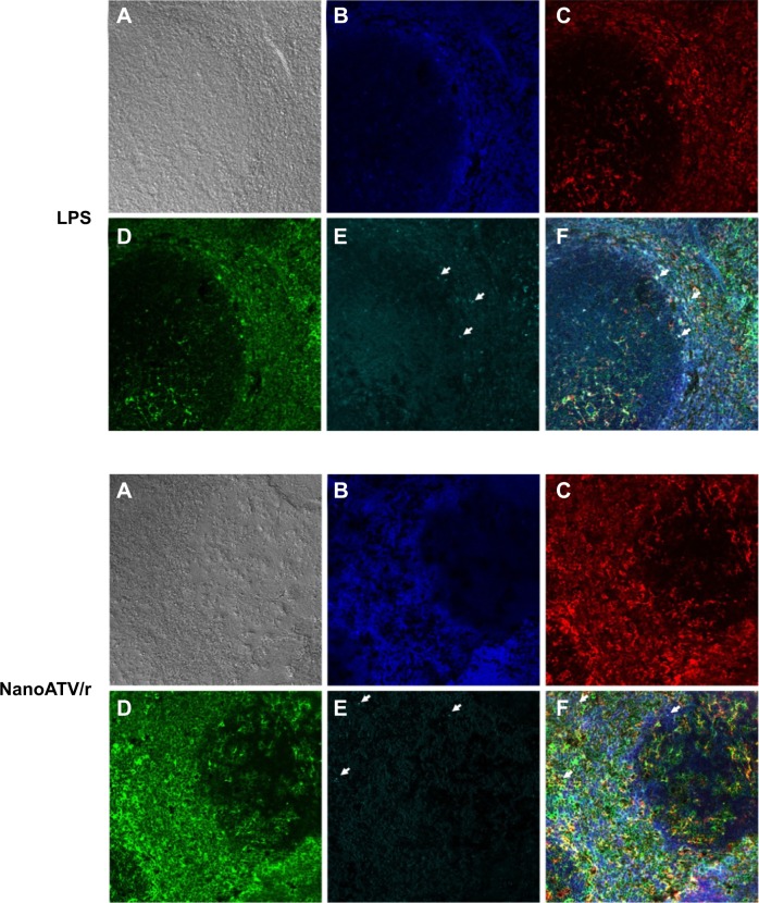
Figure 6.
Immunofluorescence staining of FAPM biodistribution in LPS-treated mouse spleen (top) or nanoATV/r treated mouse spleen (bottom).
Notes: (A) Phase contrast; (B) DAPI; (C) Iba-1; (D) folate receptor 2; (E) magnetite (arrow indicates individual magnetite); and (F) merged picture. 200×, white arrows indicate colocalized stains.
Abbreviations: FAPM, ALN-PEG-FA-coated magnetite; LPS, lipopolysaccharide; nanoATV/r, nanoformulated ritonavir (RTV)-boosted atazanavir (ATV); DAPI, 4′,6-diamidino-2-phenylindole; Iba-1, ionized calcium-binding adaptor molecule 1; ALN, alendronate; PEG, polyethylene glycol; FA, folic acid.

