FIG. 1.
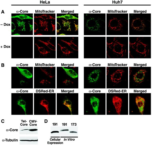
A fraction of the HCV core protein colocalizes with a mitochondrial marker in transfected cells. Confocal microscopic images of HeLa Tet-Off and Huh-7 cells expressing the core protein from a tetracycline-controlled (A) or a CMV-driven expression vector (B) are shown. Living cells were incubated with MitoTracker Red or were cotransfected with a construct expressing the ER-targeted Discosoma red fluorescent protein (DsRed-ER). Immunostaining for the core protein was performed with anti-core protein and Cy2-labeled anti-mouse antibodies 20 h after transfection. (C) Western blot analysis of core protein expressed from the tetracycline-controlled or CMV-driven vector. The blot was stripped and reprobed for tubulin as a loading control. (D) Comparison of core protein expressed from the CMV-driven vector with in vitro-synthesized truncated core protein (173 amino acids) and unprocessed core protein (191 amino acids) generated in the absence of microsomal membranes.

