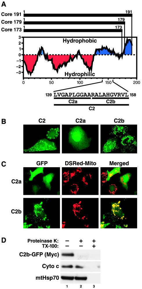
FIG. 6.
A 10-amino-acid motif functions as a mitochondrial targeting signal in the core protein. (A) Kyte-Doolittle hydrophobicity plot of full-length (Core 191) and processed (Core 179 and Core 173) core proteins. The amino acid sequences and positions of domain C2 and the subdivisions C2a and C2b within the full-length core protein are indicated at the bottom. (B) Confocal microscopy of Huh-7 cells transfected with fusion proteins of domains C2, C2a, or C2b to GFP. (C) Confocal microscopy of Huh-7 cells cotransfected with the indicated GFP fusion proteins and a construct expressing the mitochondrion-targeted Discosoma red fluorescent protein (DsRed-Mito). (D) Proteinase K digestion of crude mitochondrial fractions isolated from Huh-7 cells transfected with C2b-GFP fusion protein in the absence or presence of detergent (TX-100). A Western blot analysis was performed with antibodies against the Myc epitope to detect the Myc-tagged fusion protein, against cytochrome c (Cyto c), and against mtHsp70.

