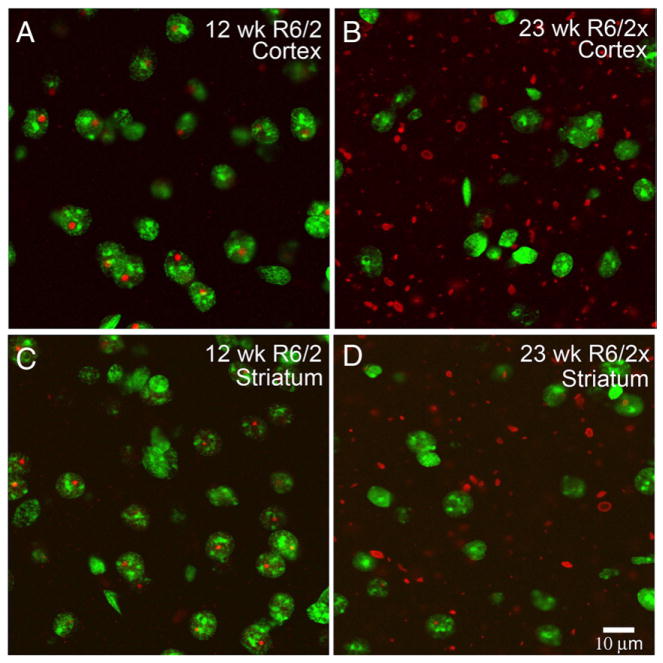
Fig. 5.
Merged CLSM images showing the location of aggregates in cortex and striatum of a 12 week old R6/2 mouse and a 23 week old R6/2x mouse, with respect to cell nuclei. Aggregates were detected by immunolabeling for the N-terminus of huntingtin using the EM48 antibody (as visualized with ALEXA594-conjugated donkey anti-mouse IgG), while neuronal nuclei were detected with the Sytox nuclear stain. Immunolabeling detects aggregated (red) mutant protein in Sytox-stained (green) neuronal nuclei in R6/2 mice but in the neuropil in R6/2x mice. These results indicate that the expanded mutant transgene protein avoids nuclear entry and predominantly forms extranuclear aggregates in R6/2x mice. Both mice were sacrificed at morbidity. Magnification is the same in all images.

