FIG. 1.

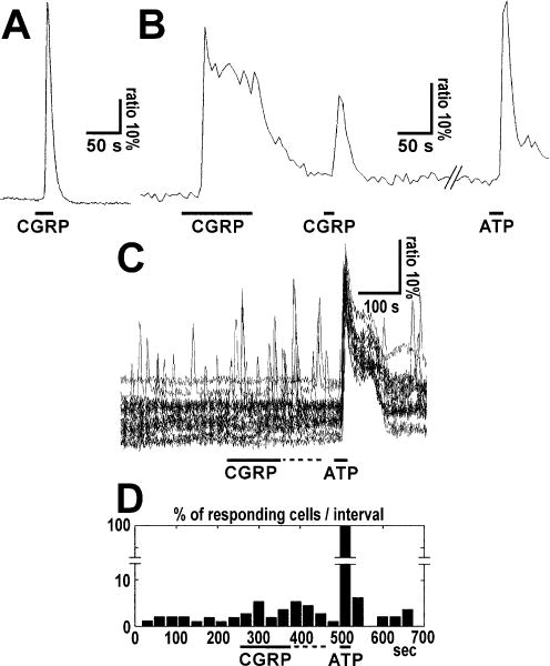
Types of calcium responses induced in cultured cerebellar astrocytes by (A) pipette application of CGRP at medium to high concentrations or (B and C) by bath perfusion at low concentrations. (A) Simple transient calcium response to CGRP application. Ratiometric Fura-2 fluorescence recordings were used to determine changes in intracellular Ca2+ concentration. Note that the onset of decay occurred in the presence of the peptide. The bar indicates local pipette application of 250 nM CGRP. (B) Peak plus sustained plateau calcium responses in a single cultured cerebellar astrocyte after CGRP application. Note that the sustained calcium plateau was maintained in the presence of CGRP and Ca2+ returned to the basal level when the peptide was washed out. The bar indicates local pipette application of CGRP (50 μM) or ATP (100 μM) as control. (C) Ratiometric traces and (D) histogram of the percentage of responding cells per time interval are shown. CGRP (1 nM) and ATP (100 μM) were applied as indicated by bars. Only cells that responded at least once to CGRP were included. In D the columns represent the percentage of astrocytes which exhibited transient calcium responses in each 30 s time interval. The statistical analysis conducted by comparing two groups of seven time intervals corresponding to (i) the total CGRP period (from 240 to 450 s, i.e. CGRP application period plus the following 90 s) and (ii) the pre-CGRP application period (from 30 to 240 s) by means of the Wilcoxon signed-rank test showed that the increase in spike frequency was significant (P = 0.0313).
