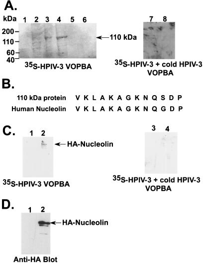
FIG. 1.
Identification of nucleolin as an HPIV-3 envelope binding protein. (A) VOPBA of A549 protein fractions eluted from the anion exchange column with 35S-HPIV-3 in the absence (lanes 1 to 6) and presence (lanes 7 and 8) of excess nonradioactive (cold) HPIV-3. The 35S-HPIV-3 interacting 110-kDa protein band is marked. (B) Comparison of the amino-terminal primary sequence of human nucleolin with the sequence of the 110-kDa protein. (C) 35S-HPIV-3 VOPBA in the absence (lanes 1 and 2) and presence (lanes 3 and 4) of excess nonradioactive (cold) HPIV-3 was performed by using anti-HA immunoprecipitated cell lysates obtained following transfection with HA-nucleolin (lanes 2 and 4) or an empty vector (lanes 1 and 3). (D) Western blot analysis of cell lysates (10 μg of protein) obtained from cells transfected with HA-nucleolin (lane 2) or an empty vector (lane 1) with anti-HA antibody.

