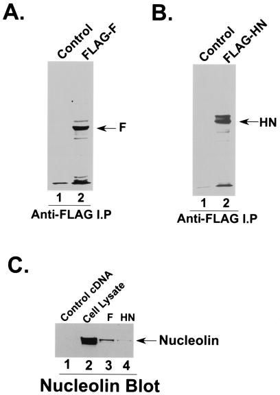
FIG. 4.
Interaction of nucleolin with HPIV-3 F and HN proteins. (A) A549 cells transfected with either an empty vector (lane 1) or HPIV-3 FLAG-F cDNA (lane 2) were pulse labeled with [35S]methionine, and the radioactive lysate was immunoprecipitated with anti-FLAG antibody prior to SDS-7.5% PAGE and fluorography. (B) A549 cells transfected with either an empty vector (lane 1) or HPIV-3 FLAG-HN cDNA (lane 2) were pulse labeled with [35S]methionine, and the radioactive lysate was immunoprecipitated with anti-FLAG antibody prior to SDS-7.5% PAGE and fluorography. (C) Lysates (100 μg of protein) obtained from A549 cells transfected with either an empty vector (lane 1), HPIV-3 FLAG-F (lane 3) or HPIV-3 FLAG-HN (lane 4) cDNA were immunoprecipitated with anti-FLAG antibody. The proteins bound to the washed anti-FLAG-agarose beads were subjected to Western blot analysis with antinucleolin antibody. An A549 cell lysate (lane 2) (20 μg of protein) served as a control.

