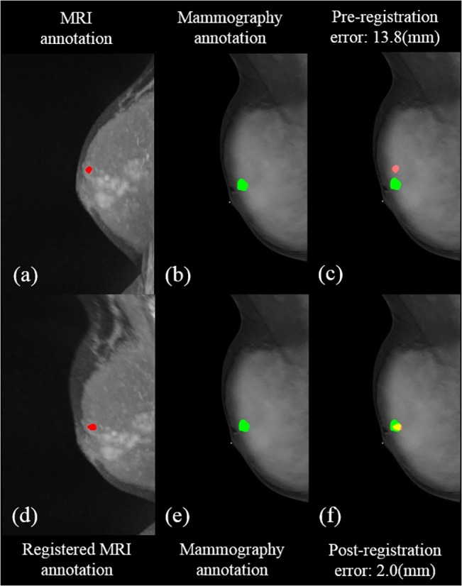
Fig 4. Illustration of the pre- and post- registration of breast MRI with annotation to fuse on mammography in a selected patient for the MLO view.

(a) the annotation (red) in MIP of MRI (b)(e) the annotation in mammography (green) (c) the fused annotations from MRI to the mammography (d) the registered MRI with annotation (red) (f) the fusion of registered annotation from MRI to mammography. The yellow area is the overlapping part between the annotations of the fused MRI and mammography.
