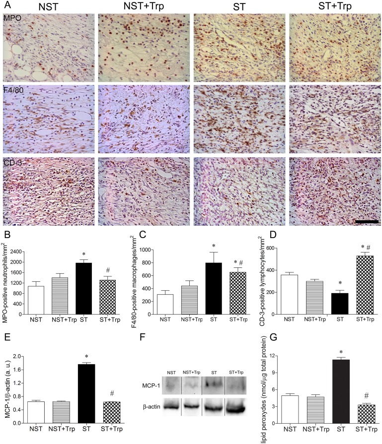
Fig 3. L-tryptophan administration reverses stress-induced alterations on inflammatory response and lipid peroxidation.
Mice were daily submitted to restraint stress and treated with L-tryptophan (Trp) or vehicle until euthanasia. Two days after the beginning of the stress protocol, two full-thickness excisional lesions (8-mm diameter) were made on the dorsal skin. Five days after wounding, lesions were collected and the number of myeloperoxidase (MPO)-positive neutrophils (B), F4/80-positive macrophages (C) and CD-3 positive T lymphocytes (D) were counted. Representative images (A) of staining for neutrophils, macrophages and T cells on wound area were presented. In addition, the protein levels of monocyte chemotactic protein-1 (MCP-1) (12 KDa) (E) were estimated in wound lysate by western-blotting. Representative images for immunoblotting for MCP-1 (F) were presented. The β-actin (42 KDa) was used as a loading control protein. Vertical black lines show non-adjacent bands from the same blot. To evaluate the oxidative damage in lipids, the levels of lipid peroxides (G) were measured in wound lysate using colorimetric assay. Data (n = 5) are presented as mean ± SEM. #p<0.05 vs. stressed (ST) group. Bar = 50 μm.

