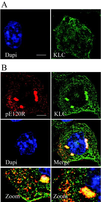
FIG. 7.
Conventional kinesin is recruited into ASFV assembly sites. (A) Distribution of kinesin light chain in noninfected cells. Noninfected Vero cells were fixed and processed for immunofluorescence by using the mouse kinesin light-chain antibody 63-90 (green). Cellular DNA was labeled with DAPI (blue). (B) Distribution of kinesin light chain in ASFV-infected cells. Vero cells were infected with the Ba71v strain of ASFV, fixed at 16 hpi, and processed for immunofluorescence. Rabbit antiserum raised against the structural protein pE120R was used to locate virus particles (red), and conventional kinesin light chain was identified by using the mouse antibody 63-90 (green). Viral DNA and cellular DNA were labeled with DAPI (blue). The bottom panels are enlarged views of the merge image. Scale bar, 8 μm.

