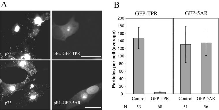
FIG. 9.
Conventional kinesin is required for anterograde transport of ASFV to the cell surface. (A) Location of ASFV particles in cells expressing GFP-TPR or GFP-5AR. Vero cells were transfected with pEL-GFP-TPR or pEL-GFP-5AR as indicated. Cells were infected with ASFV 2 h later, fixed, and processed for immunofluorescence at 16 hpi. Virus particles were identified with an antibody against p73 (left panels) and the expression of GFP-TPR and GFP-5AR was monitored by the intrinsic fluorescence of GFP (right panels). Cells expressing high levels of GFP were selected. Scale bar, 20 μm. (B) Quantification of ASFV spread to the cell periphery in cells expressing GFP-TPR or GFP-5AR. Infected cells expressing pEL-GFP-TPR or pEL-GFP-5AR were prepared as described above. The numbers of virus particles present in the cytoplasm were compared with cells (control) on the same coverslip that were infected but did not express either GFP-TPR or GFP-5AR. Results are presented as the average number of virions present in the cytoplasm per cell. Error bars indicate the standard deviation of the means. N, number of cells evaluated.

