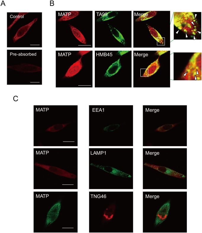
Fig 2. The MATP protein is located in the melanosomes.
(A) MNT-1 cells were stained with anti-MATP antibody before (MATP) or after antigen pre-absorbance (Pre-absorbed) for 5 min prior to adding an anti-MATP antibody. (B) MNT-1 cells were stained with anti-TA99 or-HMB45 (a melanosomal protein) antibodies together with an anti-MATP antibody. The insets are magnified, and the co-localization of MATP and each melanosomal marker is indicated by arrowheads. Scale bars = 10 μm. (C) Cells were stained with anti-EEA1, anti-LAMP1, or anti-TNG46 antibodies to detect the endosomes, the lysosomes, or the Golgi apparatus, respectively, together with an anti-MATP antibody. These representative images were captured by confocal microscopy. Scale bars = 10 μm.

