
Fig. 4.
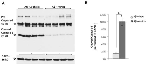
Western blot of caspase-1 shows a significant reduction in the cleaved portion of the peptide in the vinpocetine-treated group (n = 5 per group). (A) Protein lysate (including neuroretina, RPE and choroid) from Aβ + vehicle group shows intense bands at 45 kD and 20 kD, corresponding to the uncleaved (pro-peptide) and cleaved (active) forms of caspase-1, respectively. Vinpocetine markedly reduced the 20 kD band intensity consistent with a decrease in caspsase-1 activation. GAPDH (36 kD) was used to visualize protein loading levels. (B) Standard ECL method was used to quantify the 20 kD band luminescence normalized to that of GAPDH. The cleaved (active) caspase-1 peptide is reduced by greater than 80% in the Aβ + vinpo group compared to the Aβ + vehicle group. Histogram represents the mean and SEM. Statistical analysis was performed using a Mann Whitney U test, *p < 0.05.