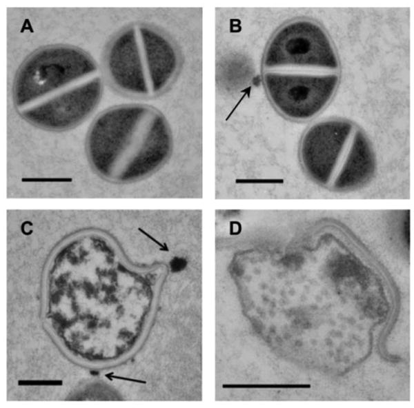
Figure 3. Curc-np induce cellular damage of MRSA.

High-power transmission electron microscopy demonstrated interaction of nanoparticles (arrows) with MRSA cells. (A) Untreated MRSA showed uniform cytoplasmic density and central cross wall surrounding a highly contrasting splitting system. (B) After 24 hours, cells incubated with control np 5 mg/ml did not exhibit changes in cellular morphology compared to untreated control. (C) After 6 hours, cells incubated with curc-np 5 mg/ml exhibited distortion of cellular architecture and edema, followed by lysis and extrusion of cytoplasmic contents after 24 hours (D). All scale bars=500 nm.
