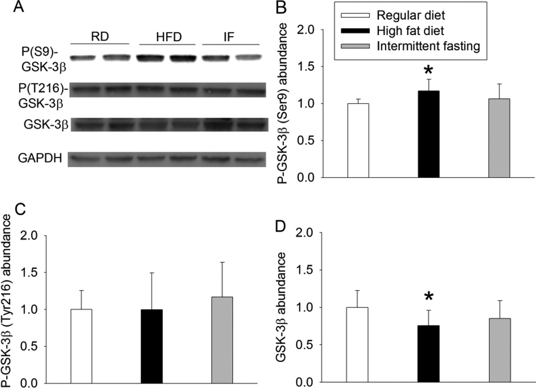
Fig. 3.
Effects of HFD and intermittent fasting on the expression of GSK-3β in the left ventricle. A: representative Western blotting. B: quantification of phospho-GSK-3β (P-GSK-3β) at Ser9. C: quantification of phospho-GSK-3β (P-GSK-3β) at Tyr216. D: quantification of GSK-3β protein. Results are means ± S.D. (n = 6 for panels B and C, = 8 for panel D). * P < 0.05 compared with control. RD: regular diet, HFD: high fat diet, IF: intermittent fasting.

