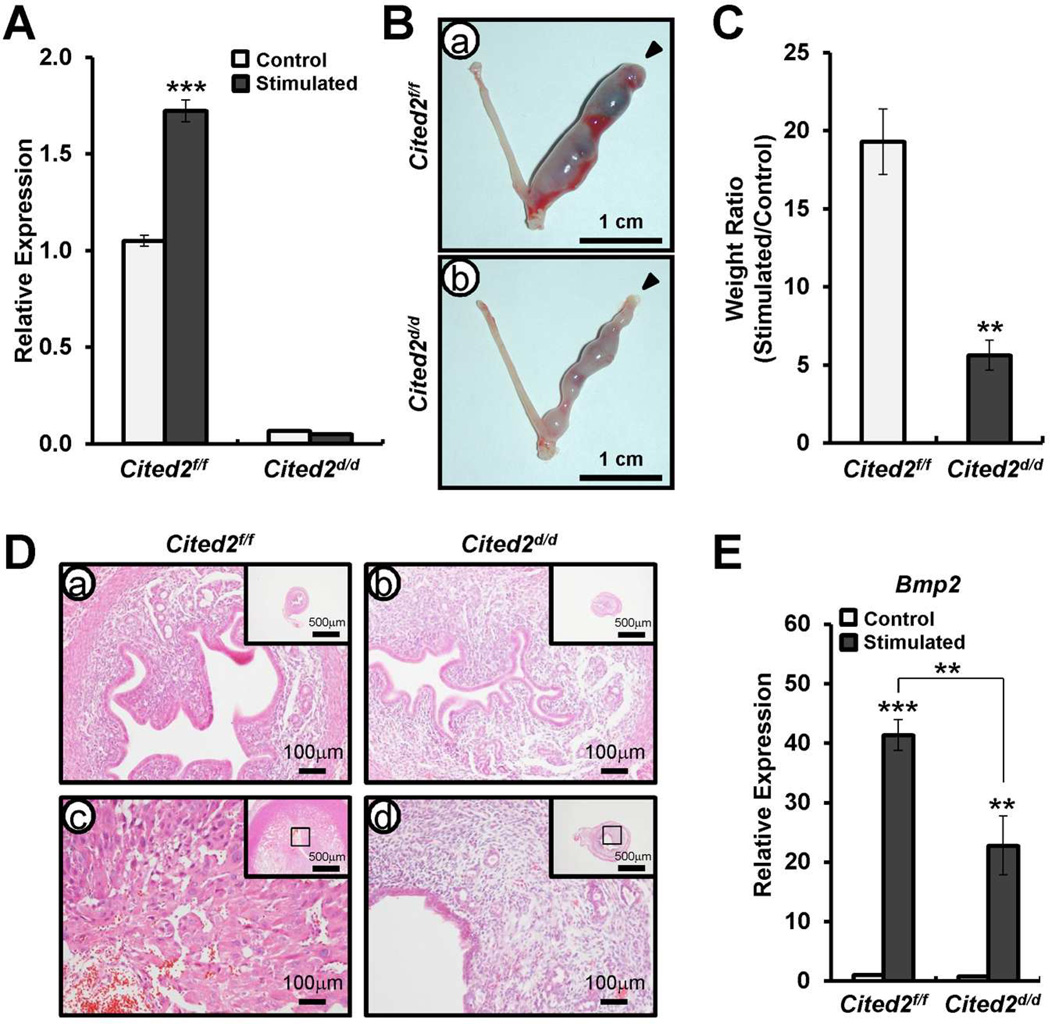
FIG. 4.
Decidual defect of Cited2d/d mice. A) Quantitative real time PCR analysis of transcript levels of Cited2 in uteri from Cited2f/f and Cited2d/d mice after artificially induced decidualization. Ovariectomized mice were primed with E2 plus P4, and one uterine horn was mechanically stimulated to mimic the presence of an implanting embryo and induce decidualization. The other horn was unstimulated and served as a control. The results represent the mean ± SEM. *** p<0.001. B) Decidualization response of Cited2d/d mice. Gross morphology of the uteri of Cited2f/f (a) and Cited2d/d (b) mice after artificially induced decidualization. C) Ratio between the weight of stimulated and control horn collected from Cited2f/f and Cited2d/d mice. Results represent means ± SEM of 3 animals per group. ** p<0.01. D) Histology of uteri was investigated by H&E staining. E) Quantitative real time PCR analysis of Bmp2 as a decidual differentiation marker in uteri of Cited2f/f and Cited2d/d mice after artificially induced decidualization. The results represent the mean ± SEM. ** p<0.01, and *** p<0.001.

