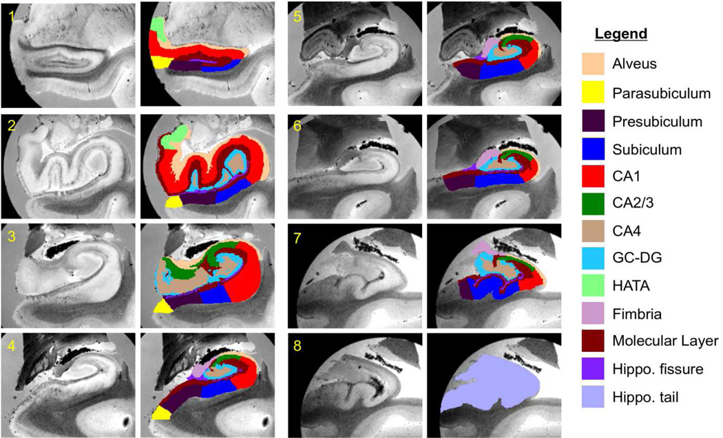
Figure 2.
Eight coronal slices from Case 14 and corresponding manual annotations. The slices are ordered from anterior to posterior. Sagittal and axial slices, as well as 3D renderings of the manual segmentation are shown in the supplementary material (Figure 15, Figure 16 and Figure 17).

