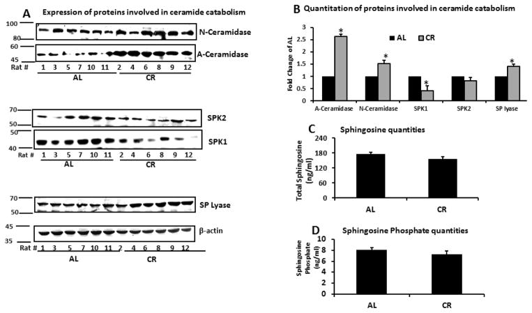
Figure 6. Expression of proteins involved in ceramide catabolism and levels of the free sphingosine and sphingosine phosphate in skeletal muscle.
A. Skeletal muscle tissue (20 mg) from rats fed either AL or CR was homogenized and expression of ceramidase, sphingosine kinase 1, sphingosine kinase 2 and SP lyase examined by standard western blotting.
B. Quantitation of western blots. All proteins standardized by β-actin.
C. Sphingosine quantities in 100 mg skeletal muscle tissue were quantified by LC-MS/MS and standardized per mg of protein.
D. Sphingosine phosphate quantities in 100 mg muscle tissue were quantified by LC-MS/MS and standardized per mg of protein.
Data are presented as mean ±SEM. *P<0.05; n=14–15, significant difference between the AL and CR groups according to a non-paired students t-test.

