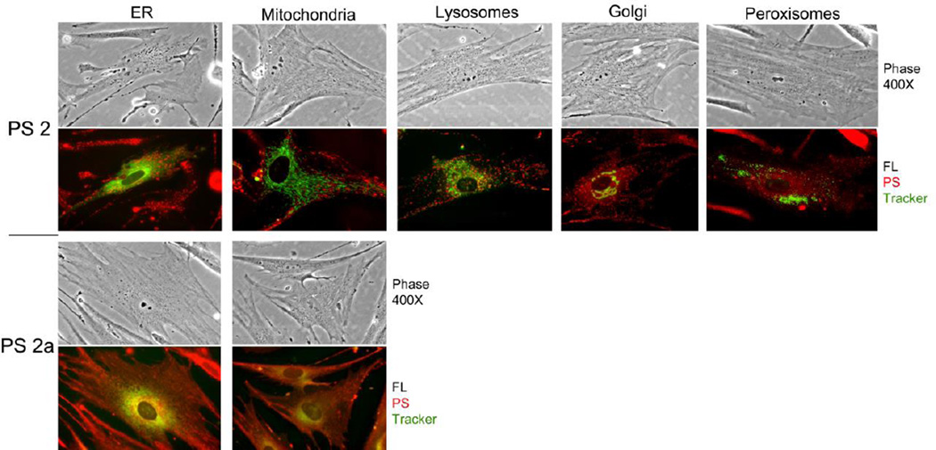
Figure 5.
Subcellular localization of compounds 2 and 2a in lung tumor fibroblasts. Primary cultures of tumor-derived fibroblasts were transduced with vectors encoding ER-GFP, Golgi-GFP or Peroxisome-RFP. After 24 h, the cells were incubated for 4 hours with compound 2 or compound 2a. Thirty min prior to the end of incubation, separate cultures were stained with Mito-tracker green or Lysotracker green. All cultures were analyzed by phase contrast and fluorescent microcopy at 400×. All organelle markers are colorized in green, and fluorescence for compounds 2 and 2a is shown in red.

