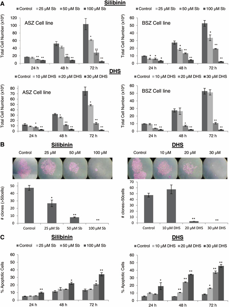
Figure 2.
The effect of silibinin and DHS on cell growth, formation of colonies and apoptotic cell death in BCC cells. (A) ASZ and BSZ cells were treated with either DMSO, silibinin (25–100 µM), or DHS (10–30 µM) for 24–72 h. After given time points, cells were collected and processed as detailed in ‘Materials and Methods’ to analyze silibinin and DHS’s effect on total cell growth in ASZ and BSZ cells. (B) ASZ cells were plated at 1 × 10⁴ cells per well and treated every 48 h with either DMSO, silibinin (25–100 µM), or DHS (10–30 µM). After 7 days, colonies greater than 50 cells were counted and the average of each group plotted. (C) ASZ cells were treated with DMSO, silibinin (25–100 µM), or DHS (10–30 µM) for 24–72 h and analyzed for apoptosis as described in ‘Materials and Methods’. Each bar represents mean ± SEM of three samples for each treatment. *P ≤ 0.05, **P ≤ 0.001, significant with respect to vehicle control.

