Figure 1.
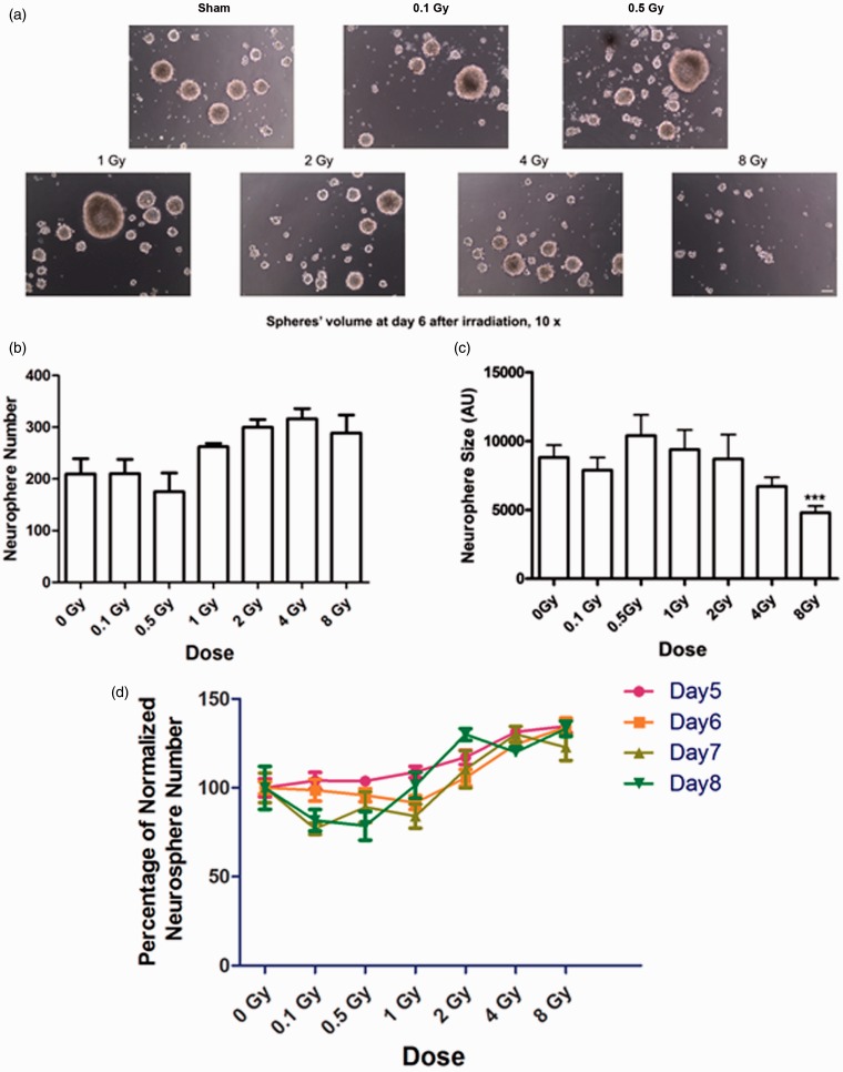
Exposure to high and low doses of 137Cs γ rays does not affect survival or self-renewal of neural stem/progenitors from the SVZ but reduces their proliferation. A single cell suspension of NSPs at second passage was exposed to γ rays in a dose range from 0 to 8 Gy and then seeded into 24-well tissue culture plate wells at 2.5 × 10⁴ cells/ml. The cells were then propagated in a biochemically defined growth medium supplemented with 20 ng/ml EGF and 10 ng/ml FGF-2 for 6 days in a humidified incubator maintained at 5% CO2 and 95% room air at 37℃. (a) Images of representative neurospheres after sham treatment or exposure to γ rays. Photographs were taken at 10 ×. Scale bar is 10 µm. (b) Average number of spheres formed per well after irradiation (n = 8) at indicated doses. There was no significant change in the number of neurospheres after irradiation compared with control (0 Gy; p > .05 by ANOVA). (c) Average size of neurospheres after irradiation at indicated doses (n > 50/group). After exposure to 8 Gy, the size of neurospheres decreased significantly (p < .001 by ANOVA). p values were determined by Kruskal–Wallis test followed by Dunn’s multiple comparisons test compared with 0 Gy. (d) Plot of neurosphere abundance normalized to control over radiation doses. There was no significant change in the number of neurospheres from 5 to 8 days after irradiation (p > .05 by repeat ANOVA). Bars represent averages ± SEM. Data represent averages of the three independent experiments. SVZ = subventricular zone; NSPs = neural stem/progenitor cells; ANOVA = analysis of variance; EGF = epidermal growth factor; FGF = fibroblast growth factor.

