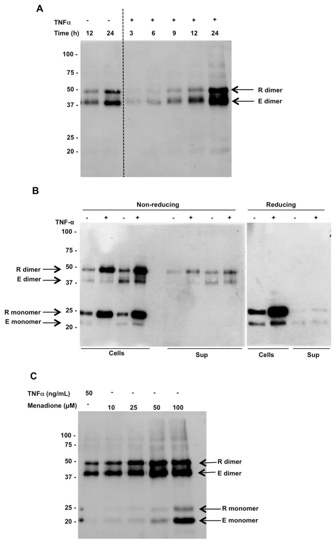
Figure 1.
Endogenous (E) and recombinant (R) Prdx2 are released from HEK 293T cells in response to treatment with TNF-α. (A) Time course of Prdx2 release in response to treatment with TNF-α analyzed by Western blotting following nonreducing SDS-PAGE of conditioned media from 293T cells expressing recombinant Prdx2 using anti-Prdx2 antibody. The two arrows indicate rPrdx2 (upper band), which is larger due to V5, and His tags and endogenous Prdx2 (lower band). (B) Intracellular and extracellular levels of endogenous (E) and recombinant (R) Prdx2 in HEK 293T cells ± TNF-α treatment for 24 h (left, gel run under nonreducing conditions; right, reducing). The dimeric form disappears under reducing conditions. (C) Release of endogenous and recombinant Prdx2 from HEK 293T cells 24 h after menadione treatment.

