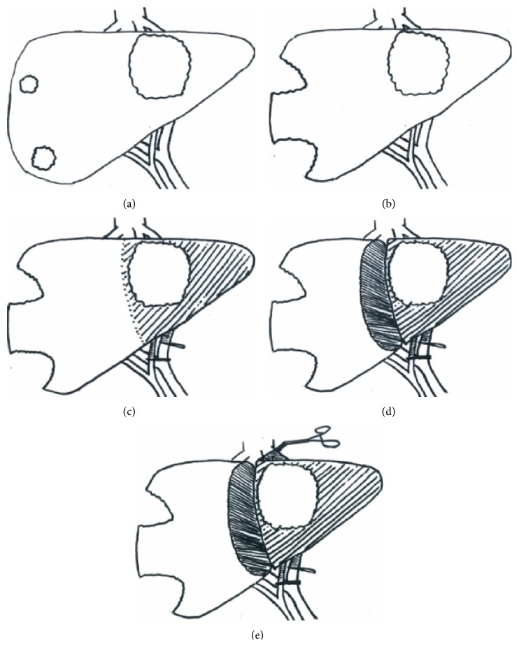
Figure 1.
Schematic diagram of ICG clearance time points in a liver with a large left-sided tumour and two small superficial right-sided tumours: (a) ICG1: preoperative, (b) ICG2: under anaesthesia following clearance of future liver remnant, (c) ICG3: during inflow control to the side to be resected, (d) ICG4: during inflow control following parenchymal transection, and (e) ICG5: the ALIIVE step, during inflow and outflow control following parenchymal transection.

