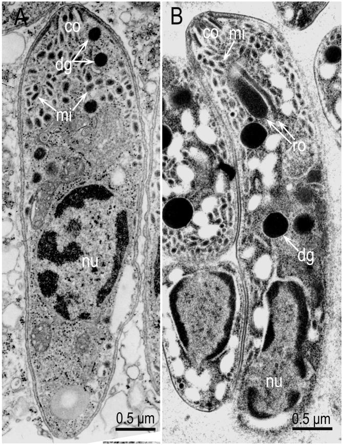
Figure 10.
Comparison of a merozoite and a bradyzoite of S. neurona. (A) Merozoite in the brain of a naturally infected horse (From Dubey et al., 1998). (B) Bradyzoite in sarcocyst in an experimentally infected cat (From Dubey et al., 2001d). Note the location of nucleus (nu), central in merozoite, and terminal in bradyzoite, and the absence of rhoptries in merozoite. Also note conoid (co), micronemes (mn) and dense granules (dg). Dense granules are often mistaken for rhoptries unless their elongated portions are visible.

