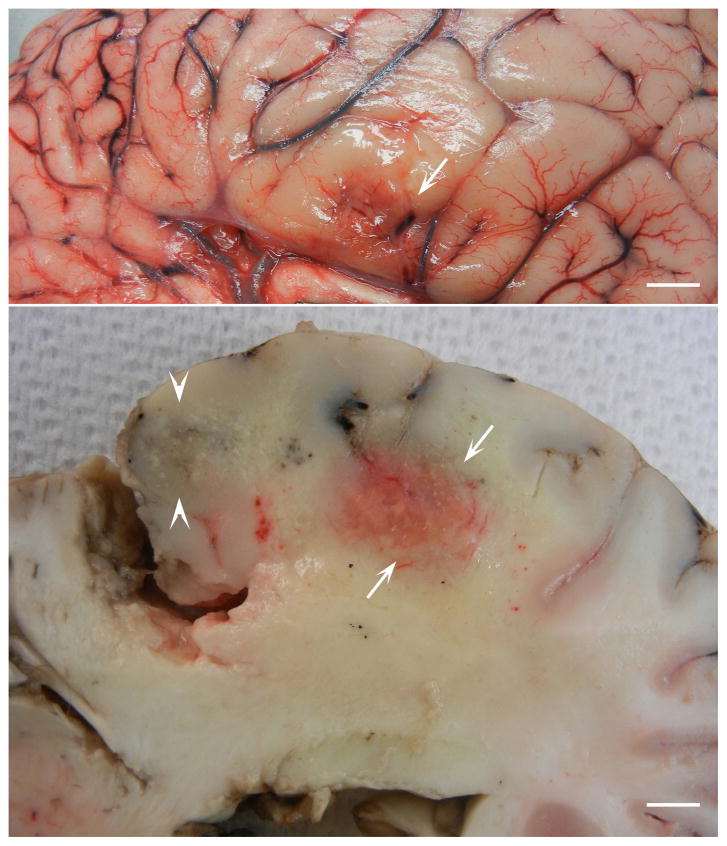
Figure 12.
Surface (top) and cut (bottom) views of cerebrum of a 20 year old Paint horse with histologically and PCR confirmed EPM. The horse had a six day history of muscle fasciculations, bruxism, difficulty eating and drinking, and circling to the left with head pressing. Note hemorrhagic and yellow discolored areas indicative of necrosis. Bar = 5 mm. (Courtesy of Uneeda Bryant).

