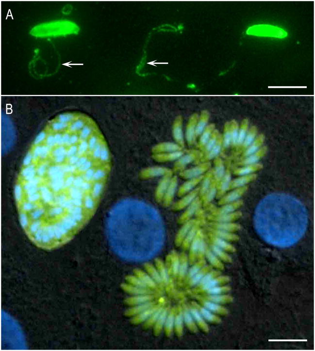
Figure 2.
Fluorescence images of S. neurona. (A) Images of gliding S. neurona merozoites stained with a monoclonal antibody (2A7-18) to the surface protein of Sn-SAG1. The formation of the trails is similar to those reported for Toxoplasma gondii. The trails are readily visualized by staining with antibodies to the major surface antigens. Gliding occurs on a variety of substrates, including coated chamber slides (50% PBS and fetal bovine serum). (B) Transgenic clone of Sarcocystis neurona expressing yellow fluorescent protein. Differential interference contrast image with epifluorescence image overlay showing a bovine turbinate cell monolayer containing a late-stage schizont and a mature schizont of a clone of S. neurona that stably expresses YFP. Host cell and parasite nuclei were stained with DAPI (blue). Bar= 10μm.

