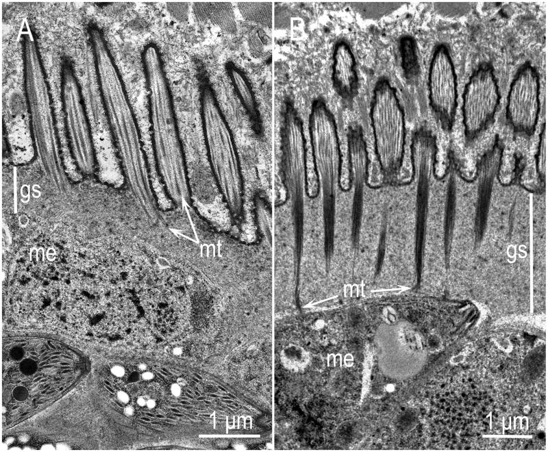
Figure 9.
Comparison of the cyst walls of S. neurona (A) and S. fayeri (B) sarcocysts by TEM. The cyst walls, including the ground substance layer (gs) of S. fayeri are thick, the microtubules (mt) are more electrondense and extend up to the pellicle of the zoites whereas the cyst walls of S. neurona are comparatively thin, the microtubules are few, and never extend deep in the gs. (From Stanek et al., 2002, and Saville et al., 2004b).

