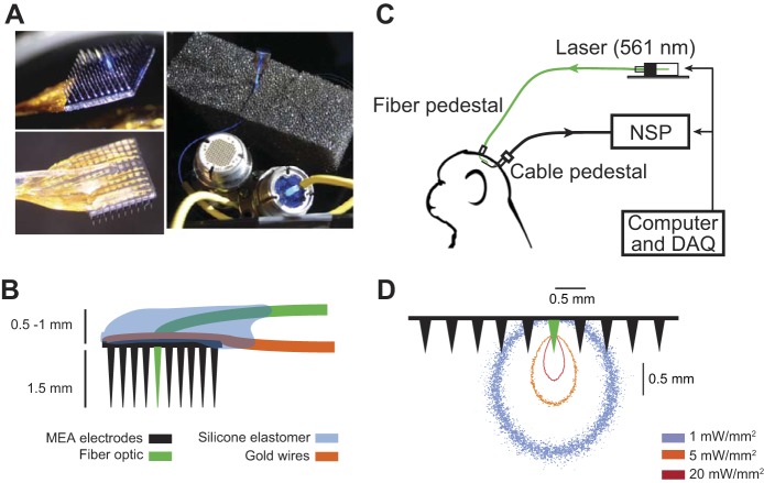
Fig. 1.
Polymer optical fiber microelectrode array (POF-MEA). A: POF-MEA viewed from the electrode side (top left), from the pad side (bottom left), and attached to two pedestals (Cereport, Blackrock Microsystems) for optical and electrical connections, respectively (right). B: schematics of the POF-MEA; an optical fiber (green) was integrated around the center of the 10 × 10 MEA. C: neural recording and optical stimulation setup. NSP, neural signal processor; DAQ, data acquisition system. D: Monte Carlo simulations of light distribution in the brain under 6-mW laser power shows isointensity contours at 1, 5, and 20 mW/mm2, respectively. (For illustration purpose, microelectrode separation, but not length, is on scale.) For details on the simulation parameters see Wang et al. (2012) and Ozden et al. (2013).

