
Fig. 7.
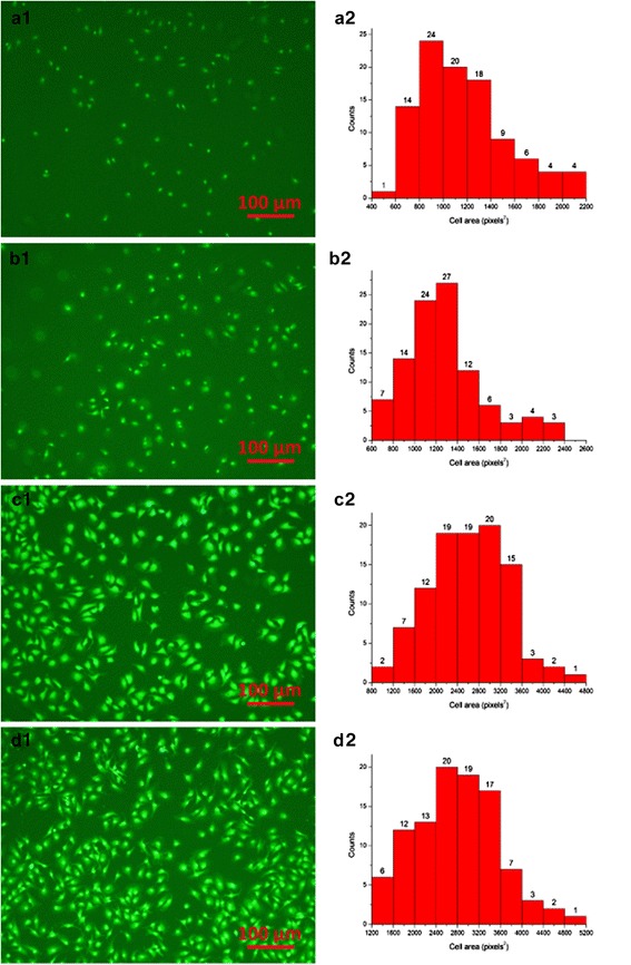
Fluorescence images and cell area distribution of ADSCs adhesion after cultivation for 5 h. a1, a2: ADSCs + PLCL; b1, b2: ADSCs + PLCL/Pluronic; c1, c2: biotinylated ADSCs + avidinized PLCL; d1, d2: biotinylated ADSCs + avidinized PLCL/Pluronic