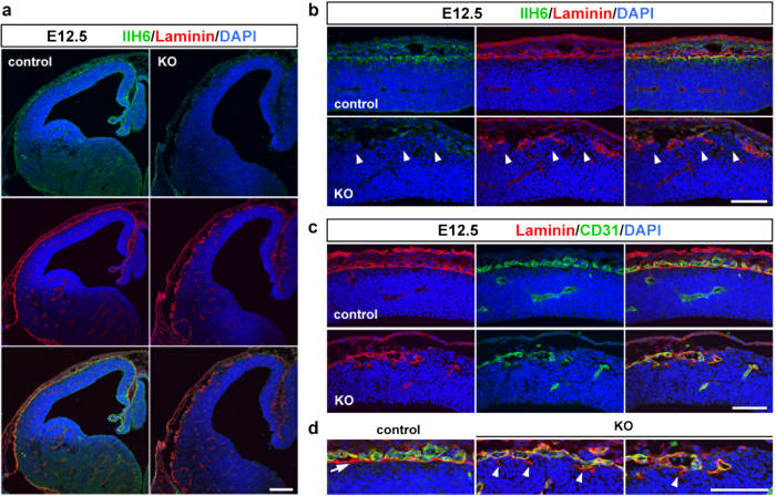
Figure 1. The Pomgnt2-KO cerebral cortex at E12.5 shows severe defects in pial basement membrane integrity.
(a) Coronal sections of the developing brain from control and Pomgnt2-KO embryos at E12.5 were immunostained with IIH6 mAb and anti-laminin pAb. (b) Enlarged images of the dorsal cortex in (a). Arrowheads indicate ectopically located cells at gaps of discontinuous laminin signals. (c) Immunohistochemistry for laminin and CD31 at E12.5. (d) Enlarged images of the pial surface in (c). The arrow indicates the laminin+/CD31− pial basement membrane in the control cortex and arrowheads show fragmented structures derived from the basement membrane in the Pomgnt2-KO cortex. Scale bars represent 200 μm (a) and 100 μm (b–d).

