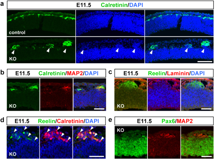
Figure 3. Abnormally located Cajal–Retzius cells and subplate neurons constitute ectopic clusters.
(a) Coronal sections of the developing brain from control and Pomgnt2-KO embryos at E11.5 were immunostained using anti-calretinin pAb. Arrowheads indicate that ectopic cell clusters present in the E11.5 Pomgnt2-KO cortex were composed of calretinin+cells. (b–e) Immunohistochemistry of the E11.5 Pomgnt2-KO cortex for calretinin and MAP2 (b), reelin and laminin (c), reelin and calretinin (d), and Pax6 and MAP2 (e). Arrowheads in (d) indicate the presence of calretinin+/reelin− cells in the ectopic cluster. Note that the MAP2+cell cluster was negative for Pax6 (e). Scale bars represent 100 μm (a) and 50 μm (b–e).

