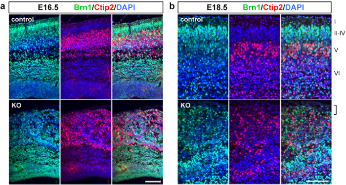
Figure 8. The Pomgnt2-KO cortex shows an abnormal cortical lamination.
(a and b) Coronal sections of the developing brain from control and Pomgnt2-KO embryos at E16.5 (a) and E18.5 (b) were immunostained for brn1 (layers II-IV) and ctip2 (layer V). The bracket in (b) shows that the most superficial region of the cortex is occupied by over-migrated neurons in the Pomgnt2-KO brain, where the cell-sparse layer I is formed in the control brain. Scale bars represent 100 μm.

