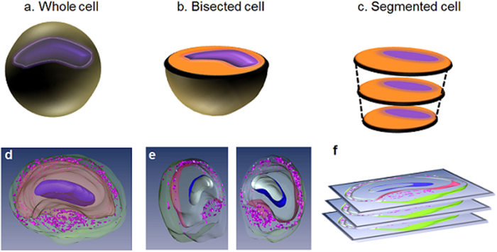
Figure 1. Schematic explaining our various sample preparation protocols.
(a) The whole cell. (b) The bisected cell to expose the intracellular organelles. (c) The segmented model for facilitating histological correlation after MR imaging. The intracellular compartments are assigned as such: nucleus (blue dots), cytoplasm (yellow areas), and dense fibers (green) around the satellite cells. 3D reconstructed image of single neurons in each sample preparation respectively d-f. The 3D visualization was generated after interpolating multiple slices of 3D MRM images in Fig. 3a. Nucleus in the cell (dark purple), cytoplasm (beige), and satellite cells (purple) in the satellite cell region (green).

